AI models improve surgical recognition of complex pelvic anatomy
Researchers have developed AI-driven surgical navigation tools to assist clinicians in identifying critical pelvic anatomy, including nerves and vessels. These systems aim to aid surgeons in navigating the complex pelvic environment and improving the recognition of key structures.

Surgical precision within the complex environment of the human pelvis is undergoing a technical shift as new modeling systems assist clinicians in identifying critical anatomical structures. Research across multiple surgical disciplines demonstrates that these tools can enhance the recognition of nerves, vessels, and organs, potentially addressing the long-standing challenges posed by anatomical variation and the narrow operative space of the pelvis.
The lateral pelvis remains a technically demanding region for colorectal, gynecological, and urological surgeons. According to findings published in npj Digital Medicine, researchers developed a model trained on 23,259 annotated images to support surgeons across various experience levels. The study observed that the assistance provided significantly improved sensitivity and specificity in identifying key structures, such as the ureter, obturator nerve, and iliac vessels, regardless of the practitioner's specialty.
Media additions
Improving Neurorecognition
One of the most significant hurdles in rectal cancer surgery is the preservation of the pelvic autonomic nervous system. Injury to these nerves can lead to persistent urogenital dysfunction, a major factor in reduced quality of life. At the Sun Yat-sen Memorial Hospital, researchers established an artificial intelligence neurorecognition system to perform real-time visual identification during surgery. By utilizing high-definition surgical screenshots and videos, the model was designed to assist surgeons in mapping the nerve course, which is often obscured by bleeding or limited visibility.
The effectiveness of this system was measured against the performance of surgeons with under 10 years of experience. While the younger surgeons required an average of 25 minutes to identify nerves in the validation set, the model completed the process in 3 minutes. Pathological examinations of tissue samples further confirmed that the structures identified by the model were indeed nerves, achieving a high rate of accuracy in distinguishing these critical components from surrounding blood vessels and membranes.
Visualization Beyond Surgery
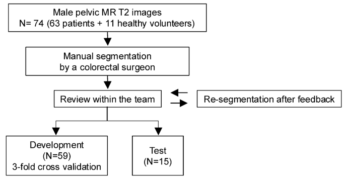
While intraoperative tools focus on real-time navigation, other developments are targeting preoperative planning. Researchers utilizing 3D MRI data from male patients have developed auto-segmentation models to delineate pelvic floor soft tissue. This approach allows surgeons to visualize individual anatomical networks before entering the operating room. Accuracy assessments showed consistent performance across different organs, though the researchers noted that the morphological characteristics of certain structures—such as the superficial transverse perineal muscle—remained more challenging for the model to segment than others, such as the internal anal sphincter.
Clinical Implications and Future Validation
The consensus among the research teams is that while these models provide substantial assistance, they are not intended to replace surgical judgment. Instead, they serve as a secondary "navigation" aid. Surgeons are encouraged to view these systems as a way to standardize care, bridging the gap between different experience levels and care environments.
| Model Focus | Key Metric | Reported Accuracy |
|---|---|---|
| Pelvic Lymph Node Dissection | Dice Similarity (Obturator Nerve) | 0.8654 |
| Male Pelvic Floor (MRI) | Dice Similarity (Internal Anal Sphincter) | 0.927 |
| Pelvic Autonomic Nerve (TME) | Precision (Nerve Category) | 0.7494 |
Despite these promising results, the path to routine clinical practice requires further validation. The authors of the npj Digital Medicine study noted that additional research using continuous intraoperative workflows is necessary to understand how these systems perform during full-length, complex procedures. Similarly, proponents of the neurorecognition system for total mesorectal excision emphasize that while the initial success in identifying autonomic nerves is notable, further research is required to definitively prove that these systems consistently lead to improved patient outcomes regarding urogenital function preservation.