Cryo-EM reveals 3D structure of dental plaque bacteria for drug design
Scientists used high-resolution imaging to visualize the 3D structure of bacteria responsible for dental plaque, revealing how they attach to host tissues. This discovery offers a potential template for developing drugs to intercept pathogens linked to periodontal disease.
Researchers have reached a new milestone in understanding the microscopic mechanics of oral health, using high-resolution imaging to map the architecture of bacteria responsible for dental plaque. This advancement offers a potential template for drug development, aiming to intercept the pathogens that contribute to periodontal disease and a range of systemic health conditions.
A multi-institutional study, published in Communications Biology, details how experts from the Okinawa Institute of Science and Technology (OIST), Tottori University, Hiroshima University, and Nagasaki University utilized cryo-electron microscopy (cryo-EM) to visualize the 3D structure of Mfa pili. These arm-like filaments allow the bacterium Porphyromonas gingivalis to adhere to host tissues and other microorganisms, a critical step in the formation of disease-causing biofilms. In Japan, the prevalence of periodontal disease is high, with approximately 80% of adults over 30 years old affected or considered at risk.
Media additions
"By understanding how P. gingivalis attaches to host tissues, establishes infection, and participates in biofilm formation, we can inform the development of future therapeutic strategies. Our detailed structural information may serve as a drug-design template for identifying compounds that block attachment and infection."
Dr. Satoshi Shibata, Lecturer at Tottori University, via Mirage News
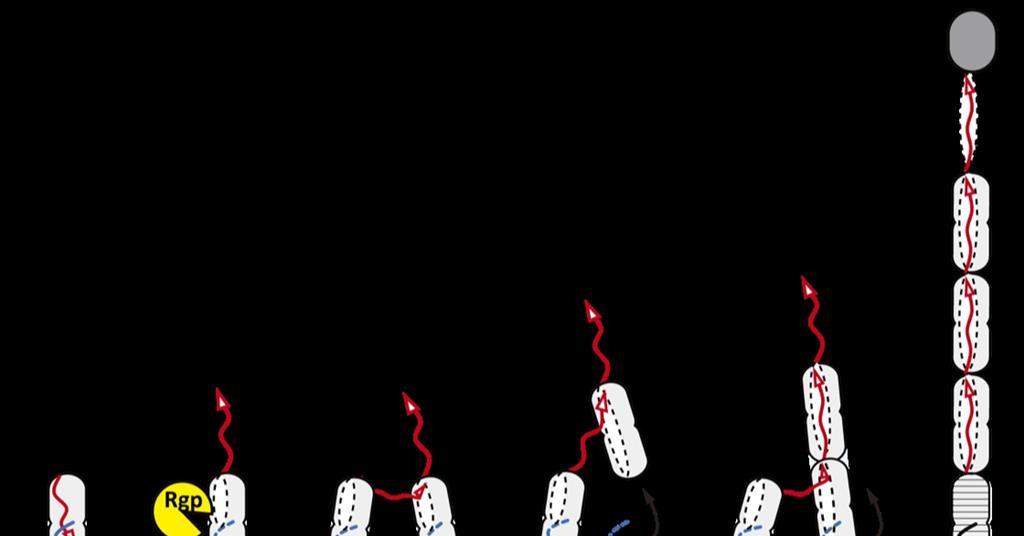
P. Gingivalis is a primary contributor to gum disease, which has been implicated in conditions such as diabetes, pneumonia, Alzheimer's disease, rheumatoid arthritis, stroke, cardiovascular disease, and adverse pregnancy outcomes. By determining the structure of Mfa1 proteins at a 3.0 Å resolution, the team identified the mechanisms that allow these filaments to assemble. Specifically, they found that the proteins Link together through a process called "strand exchange," which relies on structural stability within the C-terminus of the protein.
The research also uncovered that the Mfa filaments incorporate calcium ions, which Dr. Shibata suggests may assist the bacterium in evading the host's immune system. By combining these cryo-EM findings with computer simulations, the researchers successfully visualized how P. Gingivalis interacts with Streptococcus gordonii, another common component of dental plaque. This interaction is essential for the stabilization of the biofilm, and identifying the specific interface between these bacteria may facilitate the creation of compounds designed to disrupt the process.
The Role of Cryo-EM in Dental Research
Cryo-EM has transformed the study of oral biology by allowing scientists to bypass the limitations of traditional imaging. Unlike conventional microscopy, cryo-EM captures molecular structures at extremely low temperatures—typically between -150°C and -196°C—preserving samples in their near-natural, vitreous state. This provides researchers with a view into the dynamic ecosystem of the mouth, including the structure of dental enamel and the interfaces where biomaterials meet living tissue. The technology enables single-particle analysis for 3D reconstruction and cryo-electron tomography for the study of intact cells.
Other scientific efforts have complemented this structural work with chemical analysis. Researchers at the Hong Kong University of Science and Technology (HKUST) have investigated biofilm formation, focusing on the bacterium Streptococcus mutans, a major etiological agent of dental caries. Their work identified a small-molecule secondary metabolite, mutanofactin-697, which promotes bacterial adhesion by increasing cell hydrophobicity. While the OIST-led team focused on the physical filament structure, the HKUST team’s findings illustrate how distinct biochemical pathways contribute to the same end goal: the persistent formation of dental plaque.
Clinical and Future Implications
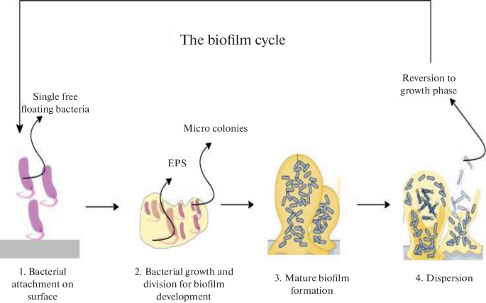
The transition from structural mapping to clinical intervention is a primary goal for the field. Dental plaque is a complex, multiphase community of bacteria, fungi, and protists shielded by an extracellular polymeric substance that can withstand stressors like extreme temperature, pressure, pH, salinity, and antibiotics. Research into the interfaces between dental biomaterials and living tissue has shown that some materials exhibit a more cohesive, seamless connection than others, which is vital for the development of improved prosthetics and implants.
Ongoing research continues to evaluate the role of phytochemicals, including flavonoids, saponins, tannins, phenols, alkaloids, and terpenoids, in the elimination of biofilms. Future work remains necessary to bridge the gap between structural findings and the clinical reduction of biofilm-related oral diseases.