Astronauts successfully conduct first diagnostic X-ray imaging in orbit
A civilian crew successfully captured diagnostic-quality X-rays in microgravity during the SpaceXray project. This milestone demonstrates new capabilities for monitoring bone health and inspecting equipment during spaceflight.
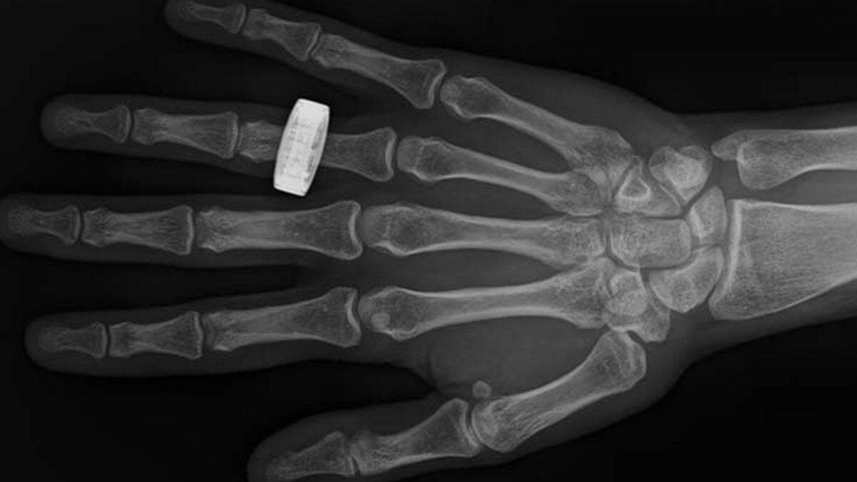
Astronauts in orbit have successfully captured diagnostic-quality X-ray images, marking the first time this medical capability has been performed during spaceflight. The achievement, which concluded a multi-year effort to adapt portable radiography for use in microgravity, signals a significant shift in how crews may monitor their health and maintain mission-critical equipment during long-duration voyages to the Moon and Mars. These findings were published on 14 July 2026 in the journal Radiology.
From Ultrasound to Digital Radiography
For more than four decades, the medical standard for in-orbit imaging has been ultrasound. While effective, ultrasound requires a medium for sound waves to travel through and relies heavily on the operator's expertise to interpret soft-tissue images. According to Sheyna Gifford, a medical doctor and assistant professor of aerospace medicine at the Mayo Clinic, relying solely on ultrasound created a gap in diagnostic coverage, particularly concerning bone injuries. Unlike ultrasound, X-rays can function in a vacuum and provide the detail necessary to assess fractures and bone density loss, which can occur at a rate of 1-2 percent per month in the hip and spine during microgravity exposure.
Related media
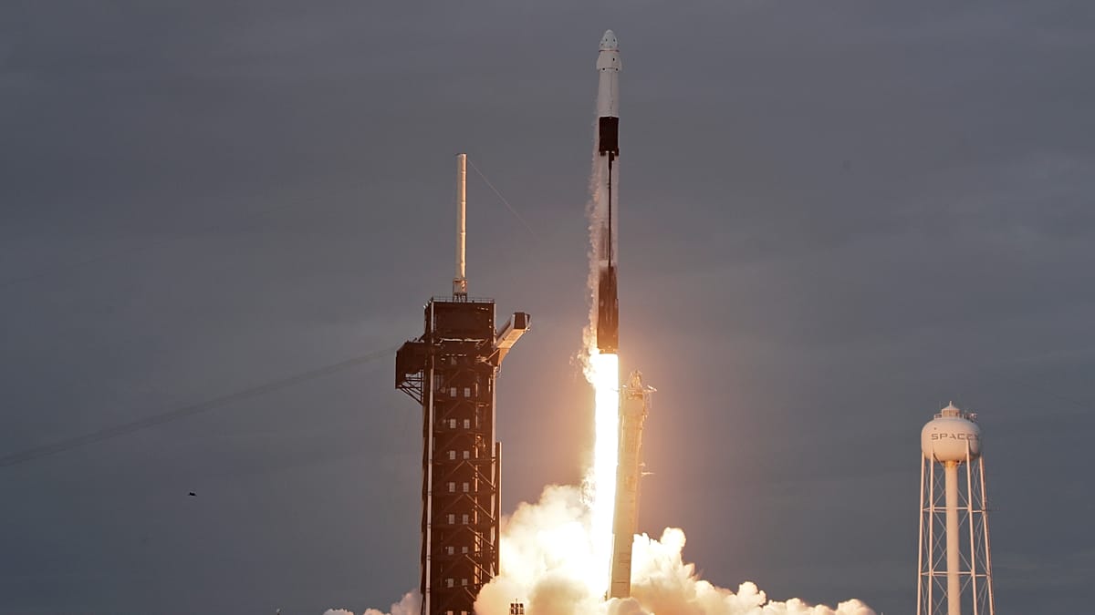
The successful test took place during the Fram2 mission, an all-civilian flight that launched on 31 March 2025. Over the course of a 3.5-day mission in polar orbit, the four-person crew utilized the Minxray IMPACT system and the Reveal 35C detector from KA Imaging to conduct imaging. The crew received only four hours of pre-flight training, demonstrating that the process could be mastered by non-medical personnel. The project, titled SpaceXray, involved collaboration between the Mayo Clinic, MIT, and the equipment manufacturers to validate that such diagnostics could be performed safely despite the radiation environment of space.
Technological Hurdles and Results
Positioning was identified as the primary challenge for in-orbit imaging. In the absence of gravity, keeping the subject, the X-ray source, and the digital detector aligned while the crew floated required significant effort. While images of extremities like hands and forearms were relatively simple to capture, central body images—such as the chest, abdomen, and pelvis—proved more difficult to align, resulting in slightly lower quality than their pre-flight counterparts. Despite this, radiologists on Earth confirmed that all captured images remained within the threshold for diagnostic use.
The equipment itself also faced rigorous real-world conditions. While the X-ray generator sustained superficial exterior damage during the flight’s return to Earth and subsequent splashdown on 4 April 2025, its internal components and output remained unaffected. This durability is crucial for future deployments where equipment may be subjected to the harsh conditions of lunar rovers or extended stays on extraterrestrial surfaces.
Broadening the Application
The utility of this technology extends well beyond human health. Researchers noted that the portability and high resolution of the systems allow for non-destructive testing of hardware. This enables astronauts to inspect electronics, spacesuits, and other mission-critical tools for hidden structural fractures or damage without needing to disassemble the items.
Experts involved in the project emphasize that these advancements also hold potential for terrestrial use, particularly in rural or remote areas where access to large-scale hospital facilities is limited. By reducing the volume and power requirements of radiography, researchers hope to eventually deploy these tools as standard equipment for both off-world exploration and global public health initiatives.
Archypedia is dedicated to independent, evidence-backed reporting. This briefing was synthesized from primary source reporting, corroborated across independent newsrooms, and verified against our Editorial Standards.
Transparency record
Evidence behind this report
This report synthesizes 12 distinct sources. Open the source ledger below to compare the underlying coverage.
- gizmodo.com
- yahoo.com
- tech.yahoo.com
- euronews.com
- minxray.com
- popsci.com
- spacexray.org
- labroots.com
- news.mit.edu
- nasa.gov
- medicaldesignandoutsourcing.com
- kaimaging.com
Prepared under the Archypedia Editorial Policy by the Niko Vale editorial desk profile. AI-assisted tools may support drafting and verification; public accountability remains with Archypedia. Report an error.