MIT engineers use mechanical stretching to control artificial blood vessel growth
Researchers are using magnetic-driven physical stimulation to mimic biological development, allowing for unprecedented control over microscopic blood vessel formation.
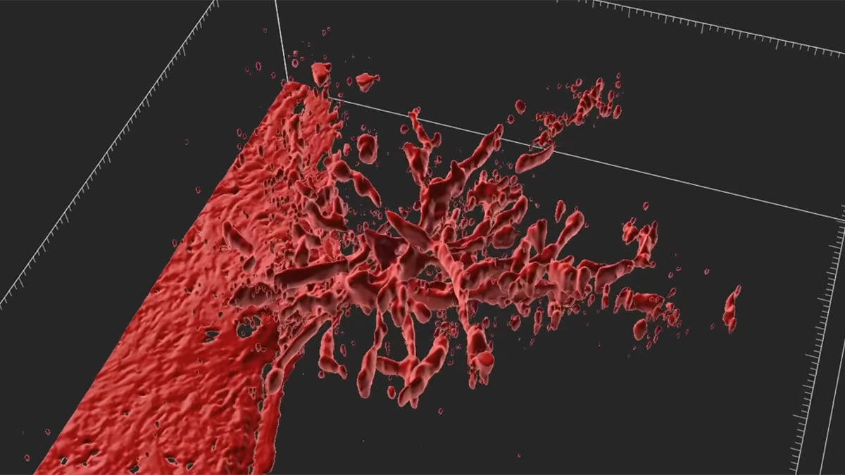
Engineers at the Massachusetts Institute of Technology have unveiled a technique to precisely control the growth of artificial blood vessels, addressing a persistent bottleneck in regenerative medicine: the creation of functional, fine-scale vascular networks within lab-grown tissues. The findings were published in the Proceedings of the National Academy of Sciences.
The core of this blood-vessel-on-a-chip
system is a platform smaller than a postage stamp. It contains a central, hollow channel lined with human endothelial cells—the cells that naturally form vessel walls—embedded within a collagen-based gel. This gel also holds a small magnet. By manipulating external magnets with a motorized three-axis system, researchers exert controlled, repetitive tension on the vessel, effectively exercising
the tissue. This physical stimulation triggers the process of angiogenesis, or the sprouting of new, microscopic capillaries.
Related media


Precision Through Mechanical Stimulation
The ability to manipulate the growth of these fine vessels is essential for the future of lab-grown organs. Without a vascular network to deliver oxygen and nutrients and remove waste, engineered tissues cannot function. Conventional methods, such as 3D printing, lack the resolution to construct delicate, thread-like capillary beds, and chemical gradients often prove difficult to control with high spatial accuracy.
According to the research team, led by Ritu Raman, an associate professor of mechanical engineering at MIT, the physical cues provided by stretching the gel allow for unprecedented architectural control. The study’s MIT co-authors include Sina Kheiri, Jessica Shah, Shashaank Venkatesh, and Roger Kamm, along with Peiyuan Chai and Ryan Flynn at Harvard University.
The team observed that the response of the cells depended on the specific parameters of the mechanical strain applied:
- Strain intensity: Stretching the gel by 5 percent of its width resulted in a higher number of new capillary sprouts.
- Vessel elongation: Increasing the strain to 15 percent produced fewer sprouts overall, but encouraged those that did form to grow significantly longer.
- Directional guidance: By changing the axis of the magnetic pull, researchers could force the growing vessels to alter their trajectory, including the creation of L-shaped structures.
The researchers noted that moving is always the takeaway of everything we do in our lab,
as physical forces play an important role in human biological development.
The Biological Link: PIEZO1
To understand why mechanical exercise triggers this growth, the researchers investigated the molecular mechanisms within the endothelial cells. The team focused on the PIEZO1 gene, which regulates ion channels that function as mechanical gatekeepers. These channels open in response to physical pressure, allowing ions to pass through the cell membrane and trigger biological changes.
By genetically editing the endothelial cells to suppress PIEZO1, the researchers observed a marked decrease in capillary sprouting despite the application of mechanical force. This confirmed that the cells interpret physical tension through these specialized ion channels, converting mechanical stimuli into internal biological instructions.
What to Watch Next
While this development offers a new layer of control for bioengineers, the researchers emphasize that they are still at an early, prototype stage. The current model lacks complex cell types, such as smooth muscle cells, pericytes, and fibroblasts, and does not yet feature continuous blood flow.
The team’s upcoming research objectives include:
- Integrating complex cell types: Expanding the system to include other essential vascular cells.
- Mimicking circulation: Introducing continuous blood flow to the chip to observe how vessels respond to fluid pressure.
- Application in muscle: Investigating whether these mechanically patterned vessel networks can improve the performance and viability of engineered muscle tissue, which has high metabolic demands.
Archypedia is dedicated to independent, evidence-backed reporting. This briefing was synthesized from primary source reporting, corroborated across independent newsrooms, and verified against our Editorial Standards.
Transparency record
Evidence behind this report
This report synthesizes 11 distinct sources. Open the source ledger below to compare the underlying coverage.
- newtoeducation.com
- news.mit.edu
- thebrighterside.news
- sciencealert.com
- meche.mit.edu
- qazinform.com
- sflorg.com
- yahoo.com
- aol.com
- industrytap.com
- drugtargetreview.com
Prepared under the Archypedia Editorial Policy by the Niko Vale editorial desk profile. AI-assisted tools may support drafting and verification; public accountability remains with Archypedia. Report an error.