Long COVID Brain Imaging Links Cognitive Decline to Potential Dementia Risk
Recent neuroimaging and cellular studies link long COVID to structural damage in the dopamine system rather than widespread brain inflammation. Researchers are now exploring whether these changes, which mirror aspects of neurodegenerative disease, could be treated with dopamine-targeting therapies.
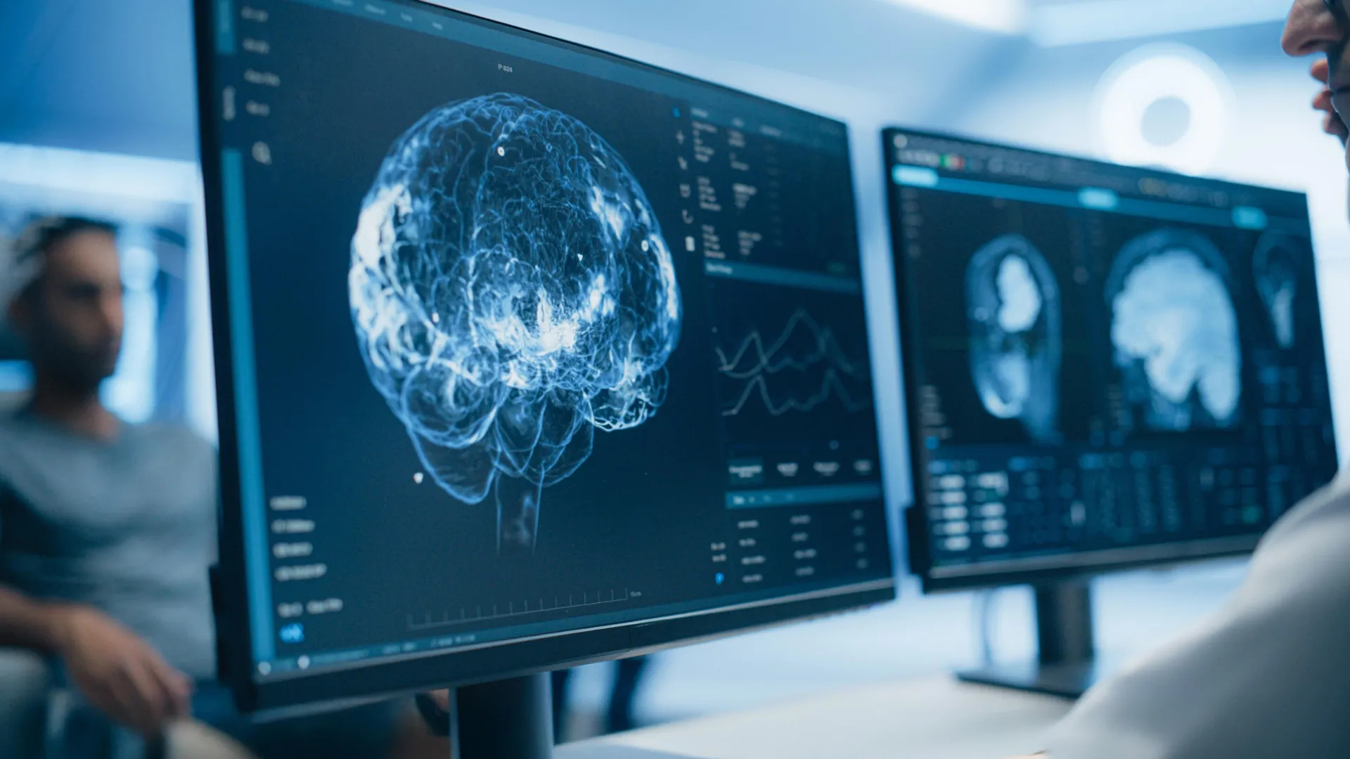
Recent neuroimaging research has shifted the scientific focus regarding long COVID from generalized brain inflammation to specific cellular and structural damage within the brain’s dopamine system. While earlier theories posited that widespread neuroinflammation was the primary driver of persistent cognitive symptoms, newer data suggest a more nuanced and localized pathology.
Imaging the Dopamine System
A study published in eBioMedicine indicates that patients with long COVID exhibit lower levels of a marker for dopamine-releasing neurons in the striatum — a brain region critical for motivation, movement, and cognition. Researchers found that this loss of nerve terminal density correlates directly with specific patient-reported outcomes: lower marker levels in the ventral striatum were linked to decreased motivation, reductions in the dorsal putamen to slowed physical movement, and decreases in the caudate putamen to memory impairment.
Related media


According to experts at the Centre for Addiction and Mental Health, this serves as the strongest biological evidence to date linking the condition to injury of dopamine pathways. This discovery provides a potential, objective basis for the debilitating fatigue and brain fog that have affected millions of individuals since the start of the pandemic.
Challenging the Inflammation Hypothesis
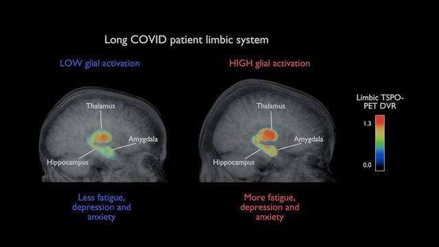
The findings regarding dopamine loss complicate the prevailing narrative that long COVID symptoms are solely the result of persistent neuroinflammation. Research conducted at the University of Turku, published in the Journal of Neurology, utilized PET imaging on long COVID patients and found no evidence of widespread inflammation in the brain. Instead, the team observed that patients with higher levels of anxiety and depression displayed increased activity in the hippocampus and amygdala, regions associated with emotional regulation and stress.
Professor Laura Airas, lead author of the study, noted that inflammation may be more prominent during the initial stages of infection but diminishes as the condition transitions into a long-term state. This suggests that for some patients, clinical approaches might need to pivot from anti-inflammatory therapies toward treatments focused on emotional regulation and stress management.
Viral Infection and Cellular Senescence
Parallel research involving lab-grown cells and autopsy samples has identified a unique biological pathway through which SARS-CoV-2 affects the brain. Studies from Weill Cornell Medicine found that the virus can infect dopamine neurons, triggering a state of senescence — where cells cease to function and begin secreting inflammatory signals. Notably, this pathway was not activated in other tested cell types, such as lung or heart tissue.
Because dopamine neuron senescence is a clinical hallmark of Parkinson’s disease, researchers suggest that long-term monitoring for movement-related neurological symptoms is warranted, even though such symptoms have not yet been widely reported in large population studies. Three existing medications, riluzole, metformin, and imatinib, have been identified as potential candidates for preventing this senescence in experimental settings, though clinical trials are required to determine if these or other dopamine-targeting drugs can effectively alleviate long COVID symptoms.
Clinical Implications and Future Directions
The convergence of these studies suggests that the neurological sequelae of COVID-19 may mirror aspects of neurodegenerative disease. A study published in Alzheimer’s & Dementia identified changes in the brain’s choroid plexus and cerebral blood flow that mirror patterns seen in early-stage Alzheimer’s disease. These structural alterations were found to be associated with cognitive impairment and specific plasma biomarkers.
| Mechanism | Impacted Regions | Clinical Symptoms |
|---|---|---|
| Dopamine nerve terminal loss | Striatum, Ventral striatum, Putamen | Lack of motivation, movement slowing, memory issues |
| Cellular Senescence | Midbrain dopamine neurons | Brain fog, lethargy, depression |
| Choroid plexus enlargement | Brain ventricles | Sleep disturbance, cognitive impairment |
| Altered neuronal activity | Hippocampus, Amygdala | Anxiety, mood changes, stress response |
What to Watch Next
- Clinical Trials: Look for updates from the University Health Network regarding trials using dopamine-modulating drugs to treat long COVID fatigue and memory deficits.
- Long-term Monitoring: Ongoing studies continue to track whether patients with long COVID demonstrate an increased incidence of Parkinson’s-related markers.
- Refinement of Biomarkers: Future research aims to establish definitive cutoff values for imaging and blood tests, potentially allowing clinicians to objectively diagnose long COVID-related brain changes in standard clinical settings.
Archypedia is dedicated to independent, evidence-backed reporting. This briefing was synthesized from primary source reporting, corroborated across independent newsrooms, and verified against our Editorial Standards.
Transparency record
Evidence behind this report
This report synthesizes 8 distinct sources. Open the source ledger below to compare the underlying coverage.
- ibtimes.sg
- medscape.com
- sciencedaily.com
- neurosciencenews.com
- insideprecisionmedicine.com
- technologynetworks.com
- thecanadianmedia.com
- news.cornell.edu
Prepared under the Archypedia Editorial Policy by the Lina Park editorial desk profile. AI-assisted tools may support drafting and verification; public accountability remains with Archypedia. Report an error.